
Techniques
Correlative imaging
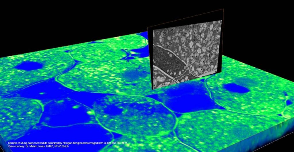
With its long-standing applications in multi-modality imaging techniques, Amira Software is the tool of choice for many researchers working with data from different imaging modalities. Amira architecture supports multi-volume data processing and visualization from the ground up. The automatic registration tools integrated into the standard edition of Amira Software with the Multi-Planar Workroom offer the flexibility of the most commonly used automated registration metrics and optimizers that allow for the adjustment of the registration parameters to meet the needs of your data.
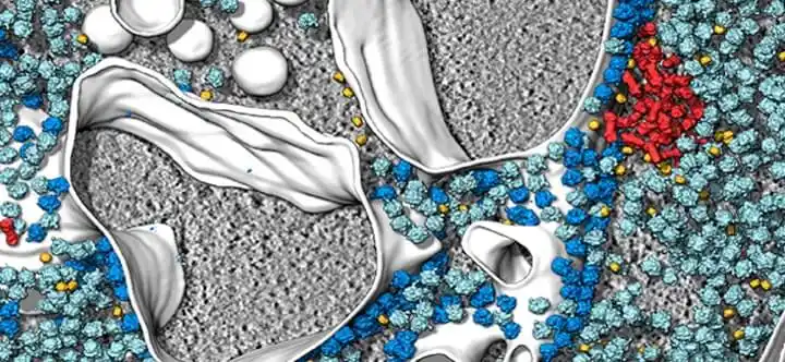
Membrane detection
Amira Software accelerates segmentation by employing automated detection of cellular features, such as filaments and membranes. By performing advanced image analysis and statistics on the cryo-tomograms, Amira Software combines data from these individual structures, averaging out noise and increasing contrast to create a composite model with higher resolution.
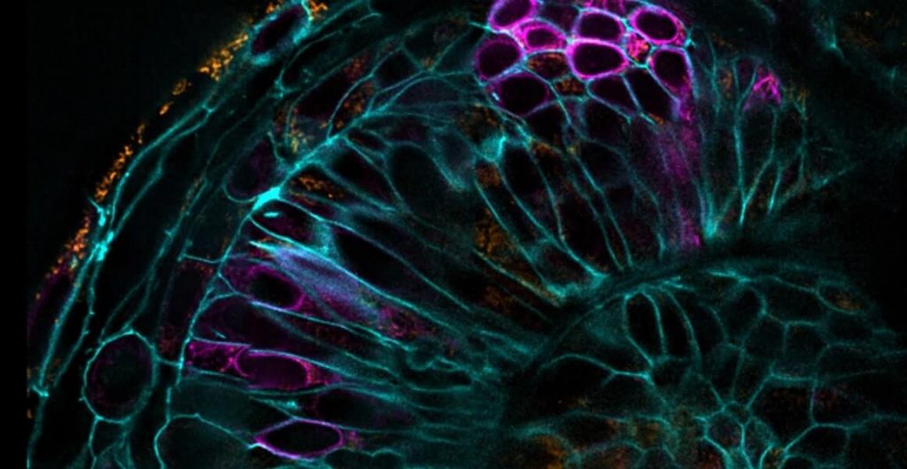
Cell detection
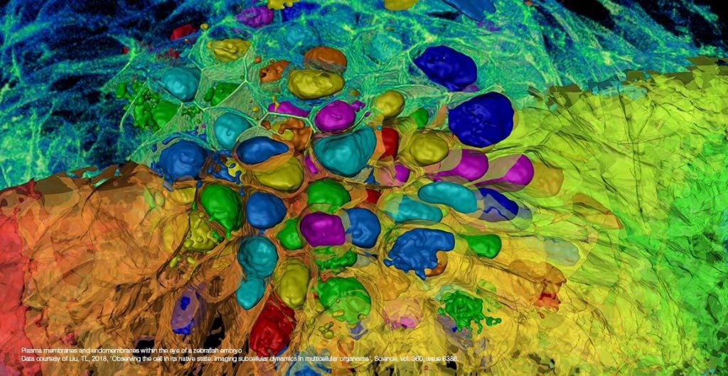
Cellular spheroids are 3D cell cultures that represent in vivo tissue better than traditional cellular monolayers, improving the ability of researchers to study cancer, diabetes mellitus, or stem cells among many applications. However, cell analysis in such 3D multicellular objects is not accurately possible, especially without destroying the object of study. Amira Software can automatically separate the cells and perform volumetric, morphometric, and intensity measurements on individual cells and their nuclei in 3D image data of these spheroids. Amira Software also helps researchers with the analysis of conventional 2D cellular monolayers.
High Content Screening
Amira Software enables multi-channel quantitivative capabilities to investigate the most demanding scientific and experimental models used in HCS, including immuno-oncology, neuroscience, and organoid health. Thanks to its incredible speed and flexibility, Amira Software helps enable advanced 2D–5D bioimaging workflows in research areas ranging from structural and cellular biology, to neuronal and spheroid morphology in 3D models. To facilitate these advanced analyses, Amira Software features deep learning for improved training and prediction, including “intelligent denoising functionality. Learn more
Multi-channel and time-series visualization
With Amira Software’s new Xplore5D extension, life scientists can easily visualize, correlate, and process large multi-channel and time series data in one platform, without worrying about image size or memory limits. With its smart file format, Xplore5D allows you to compress all imaging data without losing precious information collected during acquisition. Learn more
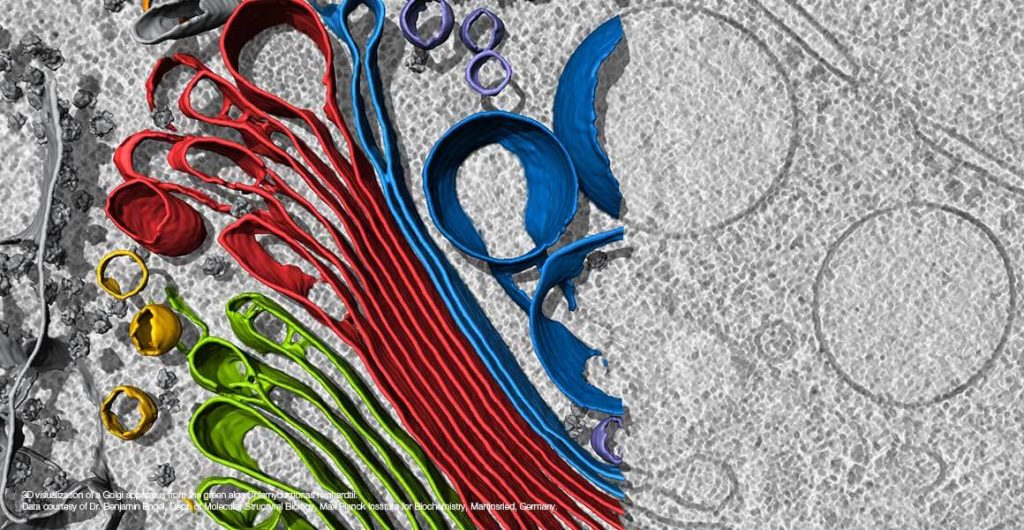
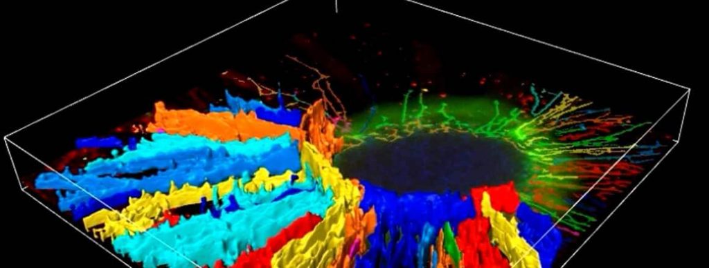
Filament tracing
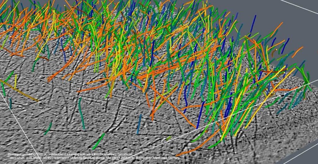
With Amira Software, you are able to use a template-matching algorithm to automatically detect and trace these fine filaments in noisy cryo-EM or DualBeam data. In addition, Amira Software enables you to reconstruct filamentous networks and edit the resulting graphs to remove image features erroneously identified as filaments or to add missing parts of a network. Graphs of filaments can then be quantified by measuring parameters such as amount, length, thickness, orientation, ranks, etc. Compelling 3D renderings can be created from the reconstructed model and the computed parameters.